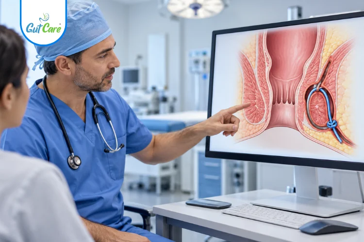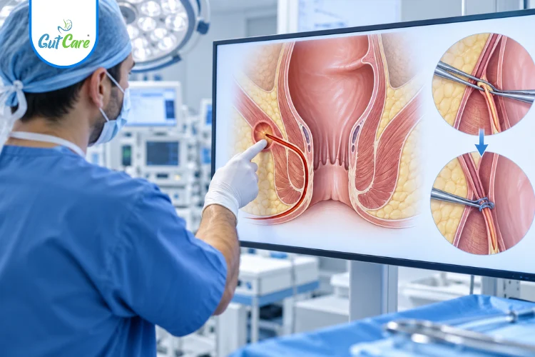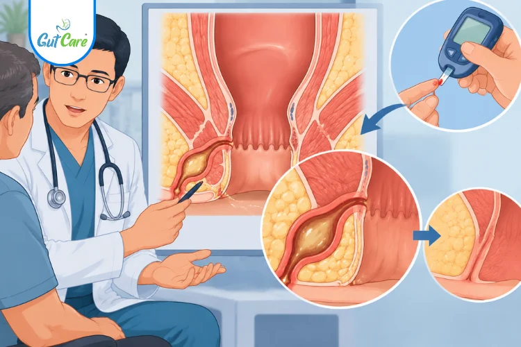
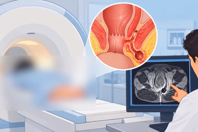
A fistula can sometimes be difficult to understand from symptoms and a physical examination alone. This is especially relevant when the fistula has a complicated path, has returned after previous treatment, or when the exact extent of the tract needs to be understood.
This is where an MRI for fistula may be recommended. An MRI can provide detailed images of the area and help doctors understand the location and course of a fistula before deciding on the next step.
At Gutcare Clinics in Bangalore, understanding the anatomy of a fistula is an important part of planning appropriate care. This guide explains why an MRI may be advised, what it can show, how the scan is performed, and what patients should know before getting one.
If you have been advised an MRI for fistula, consult a colorectal specialist to understand why the scan has been recommended and what the results may mean for your treatment plan.
Book Your Consultation Today
Why is understanding the fistula tract important?
The location and course of the fistula can influence how a doctor approaches treatment.
During an assessment, a doctor may want to understand:
- Where the fistula begins
- Where the tract travels
- Where it reaches the skin
- Whether there are additional branches
- How deep the tract extends
- Its relationship with the surrounding muscles
- Whether there are areas that require further evaluation
An MRI fistula scan can provide additional anatomical information when this level of detail is needed.
Why Is MRI Needed for Fistula?
Not every person with a fistula will necessarily need an MRI. The decision depends on the findings from the clinical assessment and the individual situation.
An MRI may be considered when the fistula’s anatomy is difficult to determine through examination alone.
When may an MRI for fistula be recommended?
An MRI may be useful in situations such as:
- The fistula appears complex
- The pathway of the fistula is unclear
- There may be more than one tract
- The fistula has returned after previous treatment
- A deeper extension needs to be assessed
- The doctor needs more detailed anatomical information before treatment
The purpose is not simply to “take a picture” of the fistula. The scan can help create a clearer map of the area so that the doctor can make a more informed treatment plan.
What Does an MRI Show in Fistula?
One of the most common questions patients ask is, “What does an MRI actually show?”
A fistula MRI can provide detailed images that help doctors assess the anatomy of the fistula and the surrounding area.
1. The fistula tract
An MRI can help identify the pathway followed by the fistula.
Instead of viewing the fistula only from its external opening, the scan can provide images that help the doctor understand what lies beneath the skin.
2. The internal and external openings
A fistula can have an opening near the anal canal or rectal area and another opening on the surrounding skin.
MRI imaging may help identify these openings and understand how they are connected.
3. The direction of the tract
The fistula may not follow a simple straight route.
MRI can help show whether the tract travels in a particular direction or follows a more complicated course.
4. Additional branches
Some fistulas may have secondary pathways or branches.
Identifying these pathways can be important when planning treatment because the visible skin opening may not represent the entire fistula.
5. Relationship with surrounding muscles
The anal region contains important muscles involved in bowel control.
An MRI can help show the relationship between the fistula tract and these surrounding structures. This information can be useful when a doctor is assessing the anatomy and considering treatment options.
6. Areas requiring further assessment
MRI images may also provide information about surrounding tissues and areas that need to be considered as part of the overall assessment.
The scan therefore contributes to a more complete anatomical picture rather than focusing only on the external opening.
MRI Fistula Mapping: Why It Matters
The term fistula mapping refers to understanding the complete pathway and anatomy of a fistula.
Imagine seeing only the opening of a road without knowing where the road goes. Treating a fistula based only on its visible opening may not provide the complete picture.
MRI can help doctors visualise:
- The starting point of the tract
- The direction of the tract
- Its depth
- Any branching pathways
- Its relationship with nearby structures
- The overall extent that needs to be considered
This information can then be considered alongside the patient’s symptoms and clinical examination.
MRI for Simple vs Complex Fistula
The need for imaging can differ depending on the situation.
A straightforward fistula may be easier to assess clinically. In other situations, the anatomy may be less obvious.
Simple fistula
When the pathway is relatively straightforward and can be adequately assessed during clinical examination, additional imaging may not always be necessary.
Complex fistula
When the tract has a more complicated course, branches, deeper extension, or a history of recurrence, more detailed imaging may be considered.
The important point is that MRI is not automatically required for every fistula. A colorectal specialist can determine whether imaging would add useful information in a particular case.
MRI Before Fistula Surgery
If surgery is being considered, understanding the fistula anatomy can be an important part of planning.
An MRI may provide information that helps the doctor understand the tract before treatment is planned.
What can the doctor understand from the scan?
The MRI findings may help answer questions such as:
- Where does the fistula start?
- Where does the tract travel?
- Is there more than one pathway?
- How deep is the tract?
- What surrounding structures are involved?
- Does the anatomy appear straightforward or complex?
The MRI report is not considered separately from the patient. The doctor combines the scan findings with the physical examination, symptoms and previous treatment history before discussing the next step.
What Happens During an MRI Scan for Fistula?
Patients often feel more comfortable when they know what to expect.
An MRI uses a large scanner to create detailed images of the area being examined. You will usually be asked to follow the instructions provided by the imaging centre before the scan.
During the scan:
- You will be positioned appropriately for the examination.
- The imaging team will explain what you need to do.
- You will need to remain still while images are taken.
- The scanner produces a series of detailed images.
- The images are reviewed and a report is prepared.
The exact preparation and scanning process can vary depending on the imaging centre and the type of examination requested.
If you have concerns about the MRI, such as discomfort in enclosed spaces, previous procedures or other medical considerations, discuss them with the imaging team beforehand.
What Happens After the MRI?
Once the MRI is completed, the images are reviewed and a report is prepared.
The next step is usually to discuss the findings with the doctor who requested the scan.
It is important not to interpret an MRI report in isolation. Medical terminology in an imaging report can be difficult to understand without the clinical context.
A colorectal specialist can explain:
- What the reported findings mean
- Whether the fistula appears simple or complex
- Whether there are additional tracts
- How the findings relate to your examination
- What treatment options may be considered
Does an MRI result decide the treatment by itself?
No single scan should be viewed separately from the overall clinical assessment.
The treatment approach depends on the individual patient’s anatomy, symptoms, previous treatment and clinical findings. The MRI is one part of that decision-making process when imaging is considered appropriate.
When Should You Discuss an MRI for Fistula With a Specialist?
If you have been diagnosed with an anal fistula, it is reasonable to ask why a particular investigation has been recommended.
You may want to discuss an MRI with your doctor if:
- The fistula has returned after previous treatment
- There is uncertainty about the tract
- More than one opening has been noticed
- The doctor suspects a more complicated pathway
- You have been advised to undergo imaging before treatment
- You want to understand what the scan is intended to clarify
The goal is not to order an MRI simply because a fistula is present. The goal is to use appropriate information to understand the condition and plan the next step.
If you have an anal fistula or have been advised to undergo an MRI, GutCare Clinics can help you discuss your symptoms, examination findings and available treatment options with a colorectal specialist in Bangalore.
Find us here:Gutcare Clinics, Indiranagar, Bangalore | Dr. Yuvraj Singh Gehlot, Digestive Health Specialist
Why Choose a Specialist Assessment for Fistula?
Fistula care involves more than identifying the external opening.
A proper assessment considers the symptoms, clinical examination, previous history and, when required, imaging findings. This approach can help create a clearer understanding of the condition before treatment is discussed.
At GutCare Clinics, patients can discuss fistula-related concerns with a specialist and understand the diagnosis, imaging findings and possible next steps in straightforward language.
5. FAQ Section
What is an MRI for fistula?
An MRI for fistula is an imaging examination that creates detailed pictures of the area around the anal and pelvic region. It may be used to understand the location and pathway of a fistula when additional anatomical information is needed.
Why do you need an MRI for an anal fistula?
An MRI may be recommended when the fistula’s pathway is unclear, appears complex, has returned after previous treatment, or requires more detailed anatomical assessment before treatment planning.
What does an MRI show in a fistula?
An MRI can help show the fistula tract, its direction, openings, possible branches, depth and relationship with surrounding structures.
Is MRI required for every fistula?
No. An MRI is not automatically required for every fistula. Whether it is useful depends on the clinical assessment and the complexity or uncertainty surrounding the fistula.
Is an MRI done before fistula surgery?
An MRI may be recommended before fistula surgery when detailed information about the fistula’s anatomy is needed for treatment planning. The decision depends on the individual case.
Can MRI detect a fistula tract?
MRI can provide detailed images that may help identify and map a fistula tract and its relationship with surrounding tissues.
What is fistula mapping on MRI?
Fistula mapping refers to understanding the course and anatomy of the fistula, including its openings, pathway, branches and relationship with nearby structures.
What should I do after getting my fistula MRI report?
Discuss the MRI report with the doctor who requested the scan. A specialist can explain the findings in relation to your symptoms and clinical examination and discuss the appropriate next steps.